The intricate folds and ridges of the human brain, unlike any other in the animal kingdom, are proving to be far more significant than previously understood. A groundbreaking new study suggests that this very complexity may be directly linked to the brain's level of connectivity and, remarkably, to our reasoning . A research team from …
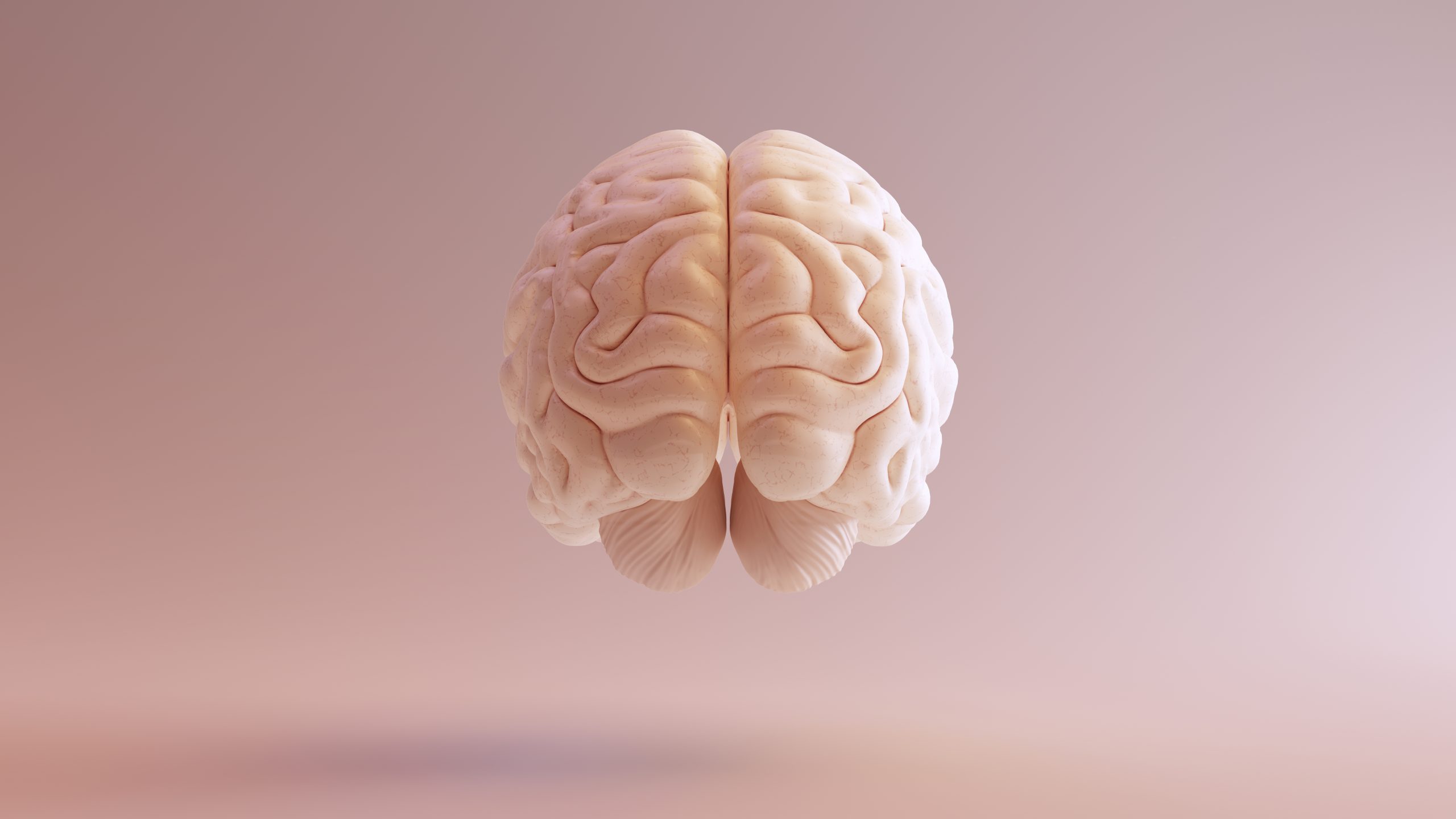
The intricate folds and ridges of the human brain, unlike any other in the animal kingdom, are proving to be far more significant than previously understood. A groundbreaking new study suggests that this very complexity may be directly linked to the brain’s level of connectivity and, remarkably, to our reasoning .
A research team from the University of California, Berkeley (UC Berkeley) meticulously examined the brain shapes and neural activity of 43 young individuals. Their focus honed in on the lateral prefrontal cortex (LPFC) and lateral parietal cortex (LPC), regions known to be critical for reasoning and high-level cognition.
The grooves and folds on the brain are scientifically termed sulci, with the smallest ones being tertiary sulci. These are the last to form during brain development, and the research team was particularly interested in how these subtle grooves might correlate with cognitive function.
(Jannsen et al., J. Neurosci, 2022)
The researchers looked at sulcal length and sulcal depth.
“The hypothesis is that the formation of sulci leads to shortened distances between connected brain regions, which could lead to increased neural efficiency, and then, in turn, individual differences in improved cognition with translational applications,” explains neuroscientist Kevin Weiner from UC Berkeley.
Their analysis unveiled a fascinating discovery: each sulcus possessed its own distinct connectivity pattern. Even more compelling, the physical structure of some of these grooves was directly tied to the level of communication between different brain areas – not just those in close proximity.
This new data builds upon the findings of a 2021 study by some of the same researchers, which had already established a link between the depth of certain sulci and cognitive reasoning. Now, we have a clearer picture of why this association might exist.
It’s astonishing to consider that between 60 and 70 percent of the brain’s outer layer, or cortex, is hidden away within these intricate folds. These patterns also undergo changes as we age, and notably, tertiary sulci can exhibit significant variations between individuals.
(Häkkinen et al., J. Neurosci., 2025)
The researchers developed models that could identify sulci with high levels of accuracy.
“While sulci can change over development, getting deeper or shallower and developing thinner or thicker gray matter – probably in ways that depend on experience – our particular configuration of sulci is a stable individual difference: their size, shape, location and even, for a few sulci, whether they’re present or absent,” notes neuroscientist Silvia Bunge, also from UC Berkeley.
This research unequivocally demonstrates that the peaks and valleys of these brain structures are far more crucial than previously understood. They are not merely random folds serving to compactly fit our brains inside our skulls; rather, they may have evolved in specific directions over time, hinting at their profound functional significance.
Looking ahead, the researchers have ambitious plans for further studying brain grooves. Ultimately, a detailed map of these sulci could potentially serve as a valuable tool for assessing brain development in children and even aiding in the early detection of neurological disorders.
However, a considerable amount of work remains before such applications become a reality. The researchers are keen to emphasize that brain fold length and depth are just two among many factors influencing our cognitive abilities.
“Cognitive function depends on variability in a variety of anatomical and functional features,” says Bunge. “Importantly, we know that experience, like quality of schooling, plays a powerful role in shaping an individual’s cognitive trajectory, and that it is malleable, even in adulthood.”
This groundbreaking research has been published in the prestigious Journal of Neuroscience.
Is the brain fog due to Iron or Menopause?
For many women, menopausal brain fog is a frustrating and pervasive symptom. While hormonal fluctuations often bear the brunt of the blame, new research is shining a spotlight on an unexpected culprit: iron. With the cessation of monthly blood loss, and thus iron loss, could rising systemic iron levels be contributing to the cognitive haze of menopause, possibly through oxidative stress?
Isn’t It Ironic?
To delve into this “iron-fog” hypothesis, researchers conducted a study involving women in early and late menopause. These participants had either low or mid-range iron levels – not deficient or anemic, but rather at the lower end of the “normal” spectrum. The study employed a multi-faceted approach, including cognitive tasks, EEGs to monitor brain activity, and MRIs to assess brain iron levels. The initial assumption was that while increased iron might enhance cognition, this benefit would be counteracted by iron-induced oxidative stress in the brain.
However, the results presented a surprising twist: higher systemic iron levels were associated with better cognitive performance and stronger EEG signals. Even more remarkably, this improvement occurred without a corresponding rise in brain iron. This suggests that in the context of menopause, iron might be a cognitive ally rather than a foe.
Rust on the Findings?
It’s important to acknowledge some limitations of this study. The COVID-19 pandemic significantly impacted recruitment, leading to a smaller sample size and persistent hesitancy towards in-person studies. Additionally, strict BMI cutoffs resulted in high exclusion rates, and hydration (a known MRI confounder) was not tracked.
Key Takeaways
Despite these limitations, this study offers crucial insights for clinicians. It underscores the importance of not dismissing iron’s role simply because ferritin (iron stores) falls within the “normal” range. Even in non-anemic menopausal women, “low-normal” iron levels may subtly impair cognitive performance.
These findings frame iron as a potentially helpful tool, rather than a double-edged sword, in managing menopausal brain fog. Clinicians should consider checking ferritin levels in all women, particularly during the menopausal transition. While specific guidelines can vary, most experts agree that a ferritin level below 30 µg/L – or even higher if symptoms are present – warrants supplementation.
Proper weight-based dosing and cofactor support can help mitigate common gastrointestinal side effects as ferritin levels are aimed to be raised to at least 100 µg/L. Larger studies are still needed to definitively confirm these findings and “iron out” the remaining details, but for many women, the fog of menopausal brain fog may already be starting to lift.
Book a Consultation
It’s easy and free!